A subscription to JoVE is required to view this content. Sign in or start your free trial.
Method Article
Fluorescence Micropipette Aspiration Assay to Investigate Red Blood Cell Mechanosensing
In This Article
Summary
The exploration of cellular behavior under mechanical stress is pivotal for advances in cellular mechanics and mechanobiology. We introduce the Fluorescence Micropipette Aspiration (fMPA) technique, a novel method combining controlled mechanical stimulation with comprehensive analysis of intracellular signaling in single cells. This technique investigates new in-depth studies of live-cell mechanobiology.
Abstract
Micropipette aspiration assays have long been a cornerstone for the investigation of live-cell mechanics, offering insights into cellular responses to mechanical stress. This paper details an innovative adaptation of the fluorescence-coupled micropipette aspiration (fMPA) assay. The fMPA assay introduces the capability to administer precise mechanical forces while concurrently monitoring the live-cell mechanotransduction processes mediated by ion channels. The sophisticated setup incorporates a precision-engineered borosilicate glass micropipette connected to a finely regulated water reservoir and pneumatic aspiration system, facilitating controlled pressure application with increments as refined as ± 1 mmHg. A significant enhancement is the integration of epi-fluorescence imaging, allowing for the simultaneous observation and quantification of cell morphological changes and intracellular calcium fluxes during aspiration. The fMPA assay, through its synergistic combination of epi-fluorescence imaging with micropipette aspiration, sets a new standard for the study of cell mechanosensing within mechanically challenging environments. This multifaceted approach is adaptable to various experimental setups, providing critical insights into the single-cell mechanosensing mechanisms.
Introduction
The unfolding discoveries in the world of cellular behaviors have accentuated the role of mechanical stimuli, such as tension, fluid shear stress, compression, and substrate stiffness, in dictating dynamic cellular activities such as adhesion, migration, and differentiation. These mechanobiological aspects are of paramount importance in elucidating how cells interact with and respond to their physiological environments, impacting various biological processes1,2.
Over the past decade, micropipette-based aspiration assays have stood out as a versatile tool in studying diverse cellular responses to mechanical stimuli. This technique offers valuable insights into the intrinsic mechanical properties of living cells at the single-cell level, including cellular elastic modulus, stiffness, and cortical tension. These assays enable the measurement of various mechanical parameters, such as cell membrane tension, pressure exerted on the cell membrane, and cortical tension (summarized in Table 1). Studying the aspirational forces has enriched our understanding of how they influence cellular functions and processes, particularly in the realm of membrane dynamics, including fragmentation, elongation, and budding3,4.
| Mechanical Parameter | Description | Seminal Approaches |
| Cell Stiffness | Measurement of a cell's mechanical rigidity and elasticity. | Aspiration of the cell membrane and analysis of deformation response to the negative pressure20,21. |
| Adhesion Strength | Evaluation of how strongly cells adhere to surfaces. | Application of controlled suction to detach adhered cells from a substrate2,22. |
| Membrane Tension | Assessment of the tension or stress within cell membranes. | Measurement of the membrane deformation in response to applied pressure23,24. |
| Viscoelastic Properties | Characterization of a cell's combined viscous and elastic behavior. | Analysis of the time-dependent deformation response to aspiration23,25. |
| Deformability | Determination of how easily a cell can change shape. | Evaluation of the extent of deformation under controlled suction20,24. |
| Surface Tension | Measurement of the tension at the cell's surface. | Assessment of the pressure required to form a micropipette membrane protrusion26. |
| Cell-Material Interaction | Study of interactions between cells and materials or substrates. | Aspiration of cells in contact with different materials and observation of interactions2,24. |
| Cell-Cell Interaction | Examination of interactions between neighboring cells. | Aspiration of a group of cells and analysis of their intercellular forces27. |
Table 1: Mechanical parameters characterized by the micropipette aspiration assay.
The micropipette-based aspiration technique has been widely used to study red blood cells (RBCs), assessing the deformability and various mechanical characteristics of RBCs, which is essential in understanding their function in the circulatory system. RBCs exhibit remarkable adaptability, preserving their mechanical versatility against deformation when navigating through the intricate capillary network and inter-endothelial clefts5,6. During this journey, RBCs must traverse through passages as narrow as 0.5-1.0 µm, subjecting themselves to a multitude of mechanical forces, including tension and compression7,8,9. They also have high sensitivity to the shear stress generated by blood flow during circulation10. These processes promote the activation of regulatory mechanisms involving calcium influx, a crucial signaling event with well-established roles in cellular responses to mechanical stimuli11,12. The complex mechanisms governing the calcium-mediated mechanosensing remain compelling subjects of ongoing investigation.
In this context, the fMPA stands as an effective approach to reveal the extent of calcium mobilization under precisely controlled mechanical forces, allowing for the simultaneous application of mechanical modulation (using the micropipette aspiration system) and visualization of calcium intensity (using fluorescent indicators). It particularly mimics the physiological scenario when the RBC travels through narrowing blood vessels. It is worth noting that the fMPA system we developed can generate pressure with a resolution of 1 mmHg. The implemented high-speed camera can achieve a temporal resolution of 100 ms and a spatial resolution at the submicron-meter level. These configurations ensure the precise application of mechanical forces to live cells and simultaneously capture the resulting cellular signaling. Moreover, due to the integrative engineered nature of this setup, the micropipette aspiration assay can be readily adapted to complement other equipment or techniques, enabling further exploration of the intricacies of cell mechanics. This versatility stands as an additional advantage of this approach.
Protocol
This protocol follows the guidelines of and has been approved by the Human Research Ethics Committee of the University of Sydney. Informed consent was obtained from the donors for this study.
1. Human RBC isolation
NOTE: Step 1.1 should be performed by a trained phlebotomist using a protocol that has been approved by the Institutional Review Board.
- Withdraw 5 mL of blood from the median cubital vein using a 19 G butterfly needle.
- Transfer the collected blood into a 15 mL tube containing 1:200 enoxaparin to prevent clotting.
- Dilute 5 µL of enoxaparin-anticoagulated blood in 1 mL of carbonate/bicarbonate buffer (C-buffer, pH = 8.5-9; Table of Materials).
- Centrifuge the diluted blood sample at 900 × g for 1 min to sediment the RBCs. Carefully decant the supernatant without disturbing the pellet.
- Perform two washes of the RBC pellet with 1 mL of C-buffer (Table of Materials), centrifuging each time at 900 × g for 1 min.
- Subsequently, wash the RBC pellet 2x with 1 mL of Tyrode's buffer using the same centrifugation conditions and then resuspend the final pellet in 1 mL of Tyrode's buffer to obtain the washed stock RBC suspension.
2. Calcium indicator loading
- Adjust the concentration of the washed stock RBC solution to 10 × 106 cells/mL in Tyrode's buffer, based on the cell count obtained using an automatic cell counter (Table of Materials).
- Label the calcium inside the RBCs by incubating with 16.67 µM Cal-520 AM, a calcium-sensitive dye, while agitating on a rotary tube mixer for 1 h.
- Dilute the RBCs in Tyrode's buffer containing 0.5% bovine serum albumin (BSA) at a 1:50 ratio. The cells are now ready for experimental use.
3. Micropipette fabrication
- Mount the borosilicate glass capillary tube (1 mm outer diameter x 0.6 mm inner diameter) onto the P-1000 micropipette puller to produce two corresponding micropipettes with closed tips at the pulling site using the preset pulling program. For this setup, use the following pulling program values: heat 516, pull 150, velocity 75, time 250, and pressure 500.
NOTE: Heating and pulling parameters set in the pulling program can be customized and are dependent on the desired settings of the experimental design12. CHECKPOINT (see Supplemental Table S1). - Open the closed tip by mounting one of the close-ended micropipettes procured after pulling onto the micropipette cutter. Adjust the heating temperature to approximately 50-60 °C.
- Locate the micropipette using a 10x eyepiece. Move the micropipette close to the borosilicate glass bead by using the knobs for adjustment.
- Change the eyepiece to 30x before positioning the micropipette as close to the borosilicate glass bead as possible without bending the pipette tip.
- Soften the borosilicate glass bead using heat by stepping onto the heating pedal. Gently insert the raw closed micropipette tip into the softened bead until the desired endpoint, the opening diameter, has been reached.
- Release the foot pedal and let the glass bead cool down. Make sure the tip of the micropipette always remains inside the bead.
NOTE: Inserting the tip further leads to larger opening diameters. - Gently extract the micropipette, leading to a clear straight cut on the closed micropipette. Confirm that the final diameter of the capillary is 1 µm.
NOTE: CHECKPOINT (see Supplemental Table S1)
4. Cell chamber preparation
- Use a diamond pencil to divide a standard 40 mm x 22 mm x 0.17 mm glass coverslip into three equal strips.
- Adhere one piece of the cut glass coverslip to the bottom of a homemade chamber holder with vacuum grease.
NOTE: The chamber holder consists of two metal (copper/aluminum) squares that are linked by a curved handle. The distance between the metal blocks must span less than 40 mm for the cut coverslip to adhere to the holder to form a parallel chamber. - Adhere the second piece of the cut glass coverslip to the top of the homemade chamber holder with vacuum grease .
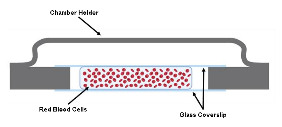
NOTE: CHECKPOINT (see Supplemental Table S1) - Inject 200 µL of the labeled RBC suspension between two coverslips using a 200 µL pipette gun (Figure 1).

Figure 1: Illustration of the cell chamber. Two cut pieces of a 40 mm x 22 mm x 0.17 mm glass cover slip are adhered to the chamber holder using grease. Between the two cut glass coverslips, approximately 200 µL of the cell solution in Tyrode's Buffer is seeded. Please click here to view a larger version of this figure.
5. Micropipette aspiration assembly
- Mount the cell chamber onto the holder stage present on the microscope platform. Adjust the position so that the cell chamber is directly above the objective (Figure 2B).
- Lower the micropipette holder to below the fluid level of the connected water reservoir.
- Inject either demineralized water or Tyrode's buffer into the fabricated micropipette and carefully remove all air bubbles using a syringe coupled with a 34 G needle (see Table of Materials).
- Unscrew the end of the micropipette holder halfway and allow the water to drip from the micropipette holder for a few seconds.
NOTE: CHECKPOINT (see Supplemental Table S1) - Insert the micropipette into the holder tip. Tighten the holder screw to ensure the micropipette is fixed.
- Insert the micropipette into the cell chamber and locate the micropipette and RBCs under the microscope. Use the micromanipulator to adjust the position.
- Lower the micropipette tip further to ensure the tip is leveled with the located RBC.
NOTE: CHECKPOINT (see Supplemental Table S1) - Zero the hydraulic pressure at the micropipette tip by adjusting the height of the water reservoir. Then, slightly raise the water reservoir to generate a subtle positive pressure at the tip.
6. Perform the fluorescence-coupled micropipette aspiration assay
- Turn on the 488 nm fluorescent excitation light source. Do not switch on the fluorescence shutter at this stage to avoid photobleaching (Figure 2C). Turn on the fluorescence camera and the transmitted camera.
NOTE: Both cameras are operated using the appropriate software (see Table of Materials). - Set up the desired exposure time (100 ms for both cameras in this study), region of interest (ROI), binning size (none for this study) for both cameras in the software. Open up the multi-dimension acquisition panel to set up the acquisition frame number, 2,000 for this study, and saving directory.
NOTE: The acquisition frame number is dependent on the desired number of aspiration events that are to be recorded. For 1 aspiration event, the range of the acquisition number should be set within 100-500, which is approximately 10-50 s. - Find the micropipette under the field of view using the micromanipulator.
- Turn on the pneumatic pressure clamp, including the control box and the clamp system (Figure 2A). Make sure the control box is in the EXTRNL mode. Compensate any offset pressure inside the system by slowly rotating the knob.
- Turn on the separate software that controls the pneumatic clamp. The software has an electrical control panel to control the discrete analog input to the clamp system. The pressure is controlled with a 20 mV/mmHg conversion factor.
- Zero the pressure inside the system. Carefully relocate the micropipette close to the RBCs. Adjust the water reservoir position until a subtle positive pressure is noticed at the micropipette tip.
- Start the acquisition in the camera-operating software. Switch on the fluorescence shutter.
- Aspirate an RBC by typing in the calculated voltage magnitude into the control panel to reach the desired pressure.
NOTE: The pressure for aspirating an RBC is typically in the range of Δp = -5 to -40 mmHg. There should be a noticeable tongue elongation within the micropipette tip (Figure 2D). - Hold the pressure for a preset period; then, release the pressure.
- Move the micropipette to pick up the next cell and repeat the experiment.
7. Fluorescence intensity analysis
- Load the saved fluorescence images into the analysis software.
- Adjust the intensity threshold using the display adjustment tab. Do this by either manually inputting the values or using the slider to ensure the fluorescence images show a clear contrast of the cell in the analysis software (see Supplemental File 1-Supplemental Figure S1).
- Scroll to the timeline at the bottom of the software. Locate the designated aspiration event.
- Click Add new surfaces. Define the analysis ROI.
NOTE: The software provides a guided five-step process to adjust and complete the segmentation (see Supplemental File 1-Supplemental Figure S2 and Supplemental Figure S3).
NOTE: Keep the ROI as small as possible to save computational resources. - Use the background subtraction slider and adjust the segmentation threshold using the slider to obtain the best segmentation outcome.
NOTE: This means that apart from the aspiration event, the background should be segmented as accurately as possible (see Supplemental File 1-Supplemental Figure S4 and Supplemental Figure S5). - Add an area filter to exclude background noises (see Supplemental File 1-Supplemental Figure S6).
NOTE: This is completed in the post process stage. - Select the statistics tab | detailed tab | average values tab. Scroll to find and select the intensity mean (see Supplemental File 1-Supplemental Figure S7).
- Export the fluorescence signal trace over time to a .csv file.
- Open the exported csv file. Subtract the background signals, Fb, from all measurements.
- Calculate the calcium intensity change, ΔFmax, using equation (1):
 (1)
(1)
Where ΔFmax is the maximum calcium intensity change, Fb, is the background intensity, and F0 is the resting intensity.

Figure 2: Fluorescence-coupled micropipette aspiration assembly. (A) An overview of the fMPA hardware system incorporating the inverted microscope combined with the brightfield and fluorescence cameras. The left side of the image depicts the homemade water manometer and the control box that allows to precisely tune the pressure of the pneumatic pressure pump. (B) The microscope stage depicting the experiment cell chamber and micromanipulator system with a single micropipette. (C) Schematic of the fMPA system setup. Concurrent imaging of brightfield (yellow) and fluorescence (blue emission, green excitation) signals utilizing two dichroic mirrors to direct the light paths from the fluorescence light source (blue) to the target, then to the cameras for imaging (green). (D) The top row depicts the brightfield images whereas the bottom row demonstrates the fluorescence images. The left represents the position of the micropipette before aspiration when the RBC is at rest.The middle column snapshots the aspiration process where the RBC experiences a negative pressure of -40 mmHg. The right depicts the cell morphology after experiencing the negative aspiration pressure. Scale bar = 5 µm. Abbreviations: fMPA = Fluorescence-coupled Micropipette Aspiration; DM = dichroic mirror; RBC = red blood cell. Please click here to view a larger version of this figure.
Results
To establish micropipette aspiration assays, we first constructed a custom cell chamber comprising two metal squares (copper/aluminum) connected by a handle. Two third-cut glass coverslips (40 mm × 7 mm × 0.17 mm) were affixed to create a chamber filled with 200 µL of RBCs suspended in Tyrode's Buffer. After introducing RBCs into the chamber, a tailored borosilicate micropipette was secured on a holder and carefully positioned within the chamber using a micro-manipulator. Subsequently, the micropipette...
Discussion
Micropipette aspiration assays embody a refined methodology, deploying substantial pressure modulation, exact spatial orchestration, and reliable temporal discernment to probe the profound intricacies of cellular biomechanics. This study places particular emphasis on the application of fMPA as a crucial tool for unveiling the nuanced mechanosensitive responses showcased by RBCs under varying stimuli. The concurrent use of brightfield and fluorescence signals enabled a multifaceted exploration of cellular phenomena, advan...
Disclosures
The authors declare that they have no competing interests to report regarding the present study.
Acknowledgements
We thank Nurul Aisha Zainal Abidin and Laura Moldovan for additional donor recruitment, blood collection, and phlebotomy support. We thank Tomas Anderson and Arian Nasser for organizing the equipment and reagents. This research was funded by the Australian Research Council (ARC) Discovery Project (DP200101970-L.A.J.); the National Health and Medical Research Council (NHMRC) of Australia Ideas Grant (APP2003904-L.A.J.); NHMRC Equipment Grant-L.A.J.; NSW Cardiovascular Capacity Building Program (Early-Mid Career Researcher Grant-L.A.J.); NSW CVRN-VCCRI Research Innovation Grant; Office of Global and Research Engagement (Sydney-Glasgow Partnership Collaboration Award-L.A.J.); L.A.J. is a National Heart Foundation Future Leader Fellow Level 2 (105863), and a Snow Medical Research Foundation Fellow (2022SF176).
Materials
| Name | Company | Catalog Number | Comments |
| µManager | Micro-Manager | Version 2.0.0 | |
| 1 mL Syringe | Terumo | 210320D | Cooperate with the Microfil |
| 200 µL Pipette | Eppendorf | 3123000055 | Red clood cell preparation |
| 22 x 40 mm Cover Slips | Knittel Glass | MS0014 | Cell chamber assembly |
| 50 mL Syringe | Terumo | 220617E | Connect to the water tower |
| Calcium Chloride (CaCl2) | Sigma-Aldrich | C1016 | Tryode's buffer preparation - 12 mM NaHCO3, 10 mM HEPES, 0.137 M NaCl, 2.7 mM KCl, and 5.5 mM D-glucose supplemented with 1 mM CaCl2. Final pH = 7.2 |
| Centrifuge 5425 | Eppendorf | 5405000280 | Red clood cell preparation |
| Clexane | Sigma-Aldrich | 1235820 | To prevent clotting of the collected blood. 10,000 U/mL |
| DAQami | Diligent | ||
| Fluorescence light source | CoolLED | pE-300 | Micropipette aspiration hardware system |
| Glass capillary | Narishige | G-1 | Micropipette manufacture |
| Glucose | Sigma-Aldrich | G8270 | Tryode's buffer preparation - 12 mM NaHCO3, 10 mM HEPES, 0.137 M NaCl, 2.7 mM KCl, and 5.5 mM D-glucose supplemented with 1 mM CaCl2. Final pH = 7.2 |
| Hepes | Thermo Fisher | 15630080 | Tryode's buffer preparation - 12 mM NaHCO3, 10 mM HEPES, 0.137 M NaCl, 2.7 mM KCl, and 5.5 mM D-glucose supplemented with 1 mM CaCl2. Final pH = 7.2 |
| High speed GigE camera | Manta | G-040B | Micropipette aspiration hardware system |
| High speed pressure clamp | Scientific Instrument | HSPC-2-SB | Cooperate with the pressure pump |
| High speed pressure clamp head stage | Scientific Instrument | HSPC-2-SB | Cooperate with the pressure pump |
| Imaris | Oxford Instruments | ||
| Inverted Microscopy | Olympus | Olympus IX83 | Micropipette aspiration hardware system |
| Microfil | World Precision Instruments | MF34G-5 | 34 G (67 mm Long) Revome air bubble in the cut micropipette and test the opening of the pipette tip |
| Micropipette Puller | Sutter instrument | P1000 | Micropipette manufacture |
| Milli Q EQ 7000 Ultrapure Water Purification System | Merck Millipore | ZEQ7000T0C | Carbonate/bicarbonate buffer & Tryode's buffer preparation |
| Pipette microforge | Narishige | MF-900 | Micropipette manufacture |
| Potassium Chloride (KCl) | Sigma-Aldrich | P9541 | Tryode's buffer preparation - 12 mM NaHCO3, 10 mM HEPES, 0.137 M NaCl, 2.7 mM KCl, and 5.5 mM D-glucose supplemented with 1 mM CaCl2. Final pH = 7.2 |
| Pressue Pump | Scientific Instrument | PV-PUMP | Induce controlled pressure during experiment |
| Prime 95B Camera | Photometrics | Prime 95B sCMOS | Flourscent imaging |
| Rotary wheel remote unit | Sensapex | uM-RM3 | Control panel for micropipette position adjustment |
| Scepter 3.0 Handheld Cell Counter | Merck Millipore | PHCC340KIT | Automatic cell counter |
| Sodium Bicarbonate (NaHCO3) | Sigma-Aldrich | S5761 | Carbonate/bicarbonate buffer preparation - 2.65 g of NaHCO3 with 2.1 g of Na2CO3 in 250 mL of Mili Q water - Final pH = 8-9. |
| Sodium Carbonate (Na2CO3) | Sigma-Aldrich | S2127 | Carbonate/bicarbonate buffer preparation - 2.65 g of NaHCO3 with 2.1 g of Na2CO3 in 250 mL of Mili Q water - Final pH = 8-9. |
| Sodium Chloride (NaCl) | Sigma-Aldrich | S7653 | Tryode's buffer preparation - 12 mM NaHCO3, 10 mM HEPES, 0.137 M NaCl, 2.7 mM KCl, and 5.5 mM D-glucose supplemented with 1 mM CaCl2. Final pH = 7.2 |
| Sodium Phosphate Monobasic Monohydrate (NaH2PO4 • H2O) | Sigma-Aldrich | S9638 | Tryode's buffer preparation - 12 mM NaHCO3, 10 mM HEPES, 0.137 M NaCl, 2.7 mM KCl, and 5.5 mM D-glucose supplemented with 1 mM CaCl2. Final pH = 7.2 |
| Touch screen control unit | Sensapex | uM-TSC | Control panel for micropipette position adjustment |
| X dry Objective | Olympus | Olympus 60x/0.70 LUCPlanFL | Micropipette aspiration hardware system |
References
- González-Bermúdez, B., Guinea, G. V., Plaza, G. R. Advances in micropipette aspiration: applications in cell biomechanics, models, and extended studies. Biophysical Journal. 116 (4), 587-594 (2019).
- Mierke, C. T. . Physics of Cancer, Volume 3 (Second Edition): Experimental biophysical techniques in cancer research. , (2021).
- Chen, Y., et al. Loss of the F-BAR protein CIP4 reduces platelet production by impairing membrane-cytoskeleton remodeling. Blood. 122 (10), 1695-1706 (2013).
- Shin, J. -. W., Swift, J., Spinler, K. R., Discher, D. E. Myosin-II inhibition and soft 2D matrix maximize multinucleation and cellular projections typical of platelet-producing megakaryocytes. Proceedings of the National Academy of Sciences. 108 (28), 11458-11463 (2011).
- Liapis, H., Foster, K., Miner, J. H. Red cell traverse through thin glomerular basement membrane. Kidney International. 61 (2), 762-763 (2002).
- Wang, H., et al. Fluorescence-coupled micropipette aspiration assay to examine calcium mobilization caused by red blood cell mechanosensing. European Biophysics Journal. 51 (2), 135-146 (2022).
- Danielczok, J. G., et al. Red blood cell passage of small capillaries is associated with transient Ca2+-mediated adaptations. Frontiers in Physiology. 8, 979 (2017).
- Diez-Silva, M., Dao, M., Han, J., Lim, C. -. T., Suresh, S. Shape and biomechanical characteristics of human red blood cells in health and disease. MRS Bulletin. 35 (5), 382-388 (2010).
- Maître, J. -. L., Niwayama, R., Turlier, H., Nédélec, F., Hiiragi, T. Pulsatile cell-autonomous contractility drives compaction in the mouse embryo. Nature Cell Biology. 17 (7), 849-855 (2015).
- Ju, L., Chen, Y., Xue, L., Du, X., Zhu, C. Cooperative unfolding of distinctive mechanoreceptor domains transduces force into signals. eLife. 5, e15447 (2016).
- Bogdanova, A., Makhro, A., Wang, J., Lipp, P., Kaestner, L. Calcium in red blood cells-a perilous balance. International Journal of Molecular Sciences. 14 (5), 9848-9872 (2013).
- . Pipette Cookbook 2018 Available from: https://www.sutter.com/PDFs/cookbook.pdf (2018)
- Cahalan, S. M., et al. Piezo1 links mechanical forces to red blood cell volume. eLife. 4, e07370 (2015).
- Sforna, L., et al. Piezo1 controls cell volume and migration by modulating swelling-activated chloride current through Ca2+ influx. Journal of Cellular Physiology. 237 (3), 1857-1870 (2022).
- High speed pressure clamp. ALA Scientific Instruments Available from: https://alascience.com/products/hspc-2sb/ (2023)
- . Teledyne Imaging Prime 95BTM Scientific CMOS Camera Datasheet Available from: https://www.photometrics.com/wp-content/uploads/2019/10/Prime-95B-Datasheet-07172020.pdf (2020)
- Lee, L. M., Lee, J. W., Chase, D., Gebrezgiabhier, D., Liu, A. P. Development of an advanced microfluidic micropipette aspiration device for single cell mechanics studies. Biomicrofluidics. 10 (5), 054105 (2016).
- Weaver, W. M., et al. Advances in high-throughput single-cell microtechnologies. Current Opinion in Biotechnology. 25, 114-123 (2014).
- Zhou, E. H., Lim, C. T., Quek, S. T. Finite element simulation of the micropipette aspiration of a living cell undergoing large viscoelastic deformation. Mechanics of Advanced Materials and Structures. 12 (6), 501-512 (2005).
- Oh, M. -. J., Kuhr, F., Byfield, F., Levitan, I. Micropipette aspiration of substrate-attached cells to estimate cell stiffness. Journal of Visualized Experiments. (67), e3886 (2012).
- Rand, R. P., Burton, A. C. Mechanical properties of the red cell membrane. Biophysical journal. 4 (4), 303-316 (1964).
- Hogan, B., Babataheri, A., Hwang, Y., Barakat, A. I., Husson, J. Characterizing cell adhesion by using micropipette aspiration. Biophysical Journal. 109 (2), 209-219 (2015).
- Henriksen, J. R., Ipsen, J. H. Measurement of membrane elasticity by micro-pipette aspiration. The European Physical Journal E. 14 (2), 149-167 (2004).
- Hochmuth, R. M. Micropipette aspiration of living cells. Journal of Biomechanics. 33 (1), 15-22 (2000).
- Pu, H., et al. Micropipette aspiration of single cells for both mechanical and electrical characterization. IEEE Transactions on Biomedical Engineering. 66 (11), 3185-3191 (2019).
- Guevorkian, K., Maître, J. -. L. Chapter 10 - Micropipette aspiration: A unique tool for exploring cell and tissue mechanics in vivo. Methods in Cell Biology. , 187-201 (2017).
- Biro, M., Maître, J. -. L. Chapter 14 - Dual pipette aspiration: A unique tool for studying intercellular adhesion. Methods in Cell Biology. 125, 255-267 (2015).
Reprints and Permissions
Request permission to reuse the text or figures of this JoVE article
Request PermissionThis article has been published
Video Coming Soon
Copyright © 2025 MyJoVE Corporation. All rights reserved